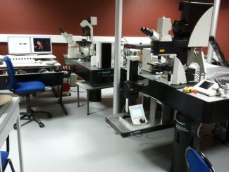
The organization of the articular collagen network was for the first time mapped specifically without any labeling thanks to the use of Second Harmonic Generation (SHG) by the imaging core facility.
A study carried out in collaboration with the laboratory, RMeS, Regenerative Medicine and Skeleton of Nantes allowed to follow the biological integration of a biodegradable scaffold made of polycaprolactone (PCL) elaborated in the form of electrospun microfibers by microscopy. This biomaterial was evaluated in a model of culture on explant but also on the animal, in a sheep model looking at the degradation of the Annulus Fibrosus (AF) during 4 weeks.
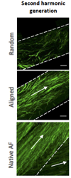
The results showed no dislocation of the implants and only one out of six samples showed partial delamination. Histological and immunohistochemical analyses revealed an integration of the implant with the surrounding tissue as well as a homogeneous organization of collagen fibers within each lamella compared to disorganized fibrous tissue and rarer in a randomly organized fibrous control scaffold .
It is the organization of the collagen network that the SHG microscopy implemented by the Imaging Core Facility of UMS2008 IBSLor has allowed to validate specifically and without labeling.
The images generated by SHG microscopy show the expression of collagen fibers and their preferential orientation within these structures.
The results thus reveal promising properties of this type of biomaterial in terms of closure of fibrous ring defects, with neotissus formation completely integrated with the surrounding ovine tissue.
Gluais M, Clouet J, Fusellier M, Decante C, Moraru C, Dutilleul M, Veziers J, Lesoeur J, Dumas D, Abadie J, Hamel A, Bord E, Chew SY, Guicheux J, Le Visage C. In vitro and in vivo evaluation of an electrospun-aligned microfibrous implant for Annulus fibrosus repair. Biomaterials. 2019 Jun ; 205:81-93.
DOI : 10.1016/j.biomaterials.2019.03.010
PUBMED : 30909111
HAL : inserm-02102713


